
TABLE OF CONTENTS
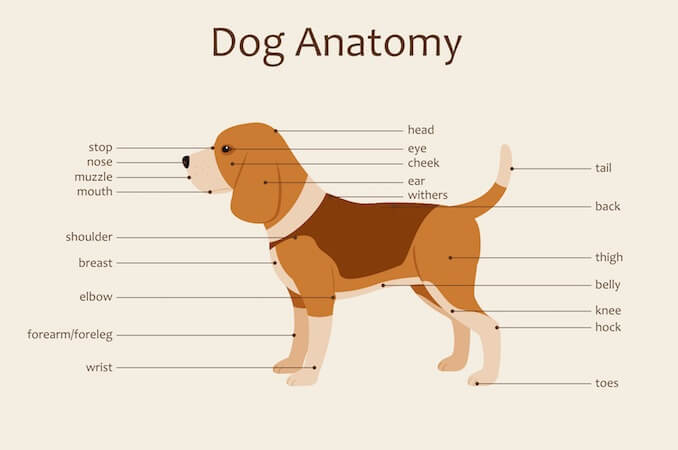
Anatomy of a Dog
Dog anatomy details the various structures of canines (e.g. muscle, organ and skeletal anatomy). The detailing of these structures changes based on dog breed due to the huge variation of size in dog breeds.
Would you be surprised to know that short dogs are more aggressive? Or taller dogs are more affectionate? It seems heavier dogs are more inquisitive and lighter dogs are more fearful. Although we should not to take such broad statements at face value, studies have shown that the size and shape of a dog can impact their behavior.
This makes us want to learn more about the anatomy of our four-legged friends. For that reason, we have put together a handy guide with some interesting facts and diagrams.
Dog Skeleton Anatomy
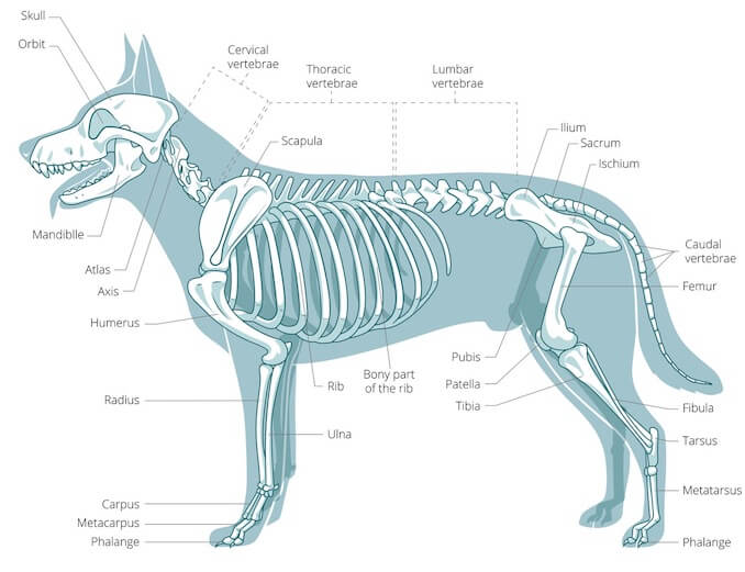
With the large range of breeds and dog sizes, despite their difference in appearance, it might be surprising to hear dog anatomy is generally the same with regards to physical anatomy and characteristics.
Dogs have a skeletal system. However, dogs don’t have a collar bone, unlike humans; providing a larger stride for running.
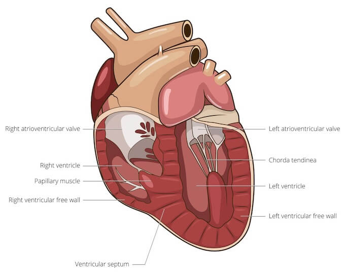
A cardiovascular system; they need powerful muscles for movement.
They have a brain for learning and teeth for eating, holding and chewing!
Despite their similarities, toy breeds have a skeleton that will mature in around 6 months. Whereas giant breeds can take between 18 months and 2 years for their growth plates to fuse.
Speaking of skeletons, a dog has 320 bones in their body (depending on the length of their tail) and around 700 muscles.
Muscles attach to bones via tendons. Depending on the breed of dog, they will have different types of muscle fibers.
You’ve probably heard about slow and fast twitch muscle fibers before.
A lurcher will have more fast twitch (anaerobic) fibers in their legs than the Alaskan Malamute who has more slow twitch (aerobic) fibers.
Lurchers have been bred for speed and agility – they require short bursts of energy or speed. Ask any greyhound, whippet or lurcher owner and they will fondly recall the crazy 5 minute runs around the yard.
Sled dogs on the other hand need continued blood flow to their muscles for endurance. This could go someway to explain why sled dogs are renowned for being “fatigue-proof!”
On the subject of leg muscles, let’s have a look at the leg as a whole in a little more detail.
Dog Leg Anatomy
Two thirds of a dog’s body weight is carried on their front legs. Only one third is carried on their hind legs.
However, the muscles on their hind legs are larger and therefore stronger.
It’s helpful to understand the anatomy of your faithful friend’s legs to identify weaknesses or injuries. It makes it much easier to explain to the vet; if you have an idea of where the problem may be.
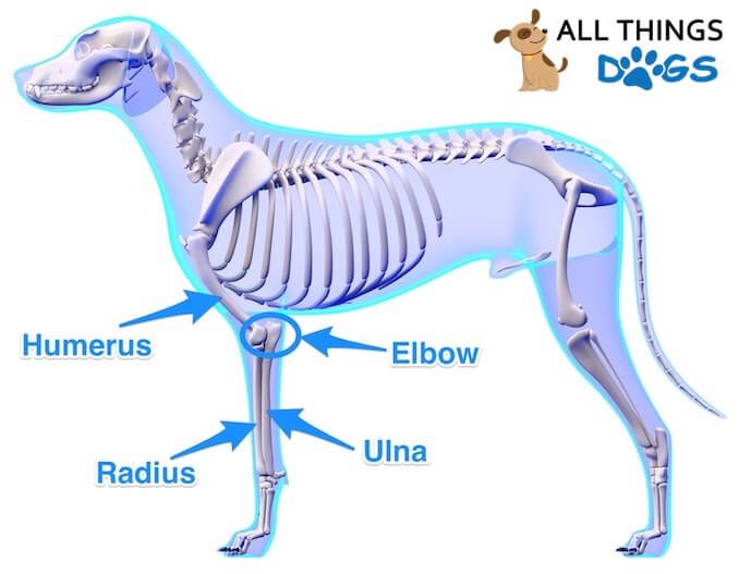
The foreleg consists of a shoulder, elbow, ulna, humerus radius and wrist.
Many large breeds can suffer with elbow dysplasia; where there is abnormal development in the joint. The most common symptom is lameness. Lesions within the elbow joint often start in puppy hood which is why it’s so important to be mindful of over-exercise!
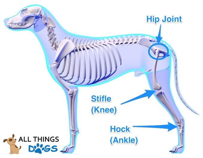
The hind leg can be confusing to some owners, but it has some of the same features as a human.
The bone between the hip and knee is the femur. Below the knee is the tibia and fibula.
Then we get to the hock; you’ve probably heard this mentioned more in horses.
The hock is like the human ankle. As with the elbow, many large breeds suffer with abnormal development in the hip joint; known as hip dysplasia. Again, lesions can start in puppy hood.
Hip dysplasia is often hereditary which is why reputable dog breeders score the hips of their dogs.
There are also environmental factors which can influence the development of hip dysplasia including: rapid weight gain/growth, diets with high calcium and Vitamin D content, over-exercise as a puppy and climbing stairs and access to slippery floors.
Still on the subject of skeletons, let’s have a look at the tail.
Dog Tail Anatomy
The tail isn’t just something which wags to show you they’re happy – it serves a much bigger function.
They can be long, short, curly or flat!
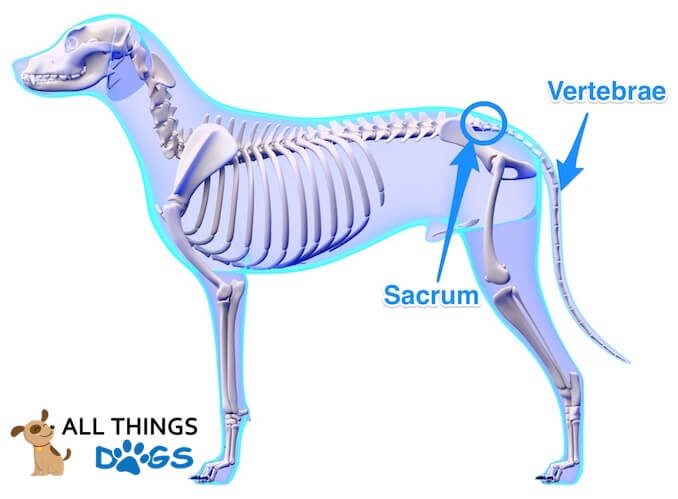
The tail is an extension of the spine, so any injuries to the tail can be quite serious.
The bones of the tail are called vertebrae, just like in the spine, and they too have discs to cushion the gap between.
The muscles and nerves found in the tail contribute to bowel control and movement which is why if a dog ever traps their tail in a door one of the first questions the vet will ask is about their toileting.
Happy Tail Happy Dog, Right?
There is a condition ironically called happy tail. This is where the dog continuously wags their tail; hitting it on anything they come into contact with. This can lead to lesions, which due to the nature of the problem (constant wagging), don’t heal. Bandaging the tail often helps and keeping the dog in large open plan areas.
Tail docking is common practice in working dogs; yet more and more research is showing that tail docking is linked to subsequent chronic pain and heightened pain sensitivity.
A dog’s tail serves many functions including counter-balancing the weight of the body when turning at speed. It is also a crucial piece of the puzzle when observing their body language.
Dog Paw Anatomy
Moving down the leg; after the hock we get to the paw, which as we know is their foot.
Their front and rear paws are very similar, just have different names.
From their hock (tarsal) joint, there are metatarsal bones which lead to their toes. In the dog’s front legs, from the carpal (wrist) joint, there are metacarpal bones which lead to their toes.
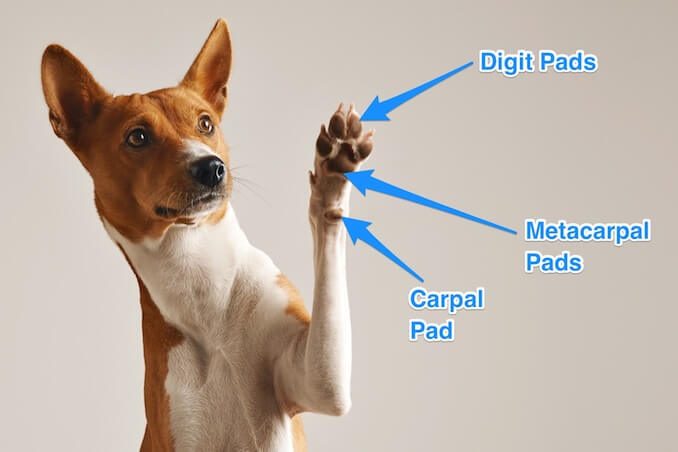
The four oval shaped pads are known as digit pads, whereas the large pad in the middle is known as either the metacarpal or metatarsal pad, depending on whether it’s their front or rear leg.
You will also notice a pad slightly further up their front leg; often sticking out. This is known as the carpal pad.
Paw pads are crucial for cushioning of the bones, providing traction and abrasion resistance. If there is trauma or injury to any one of the pads, it can often result in loss of limb function.
Aiding traction also are the dew claws; these are what you could call thumbs in human terms.
They are found higher up than the other toes and don’t touch the ground when your dog is walking.
Some dogs only have dew claws on their front legs, some on all four legs – some don’t have any. If you ever watch a dog scrambling up a steep bank, you may notice they spread their toes and use their dew claw for grip.
Some working or hunting dogs have their dew claws removed when they are puppies if it is deemed they may suffer a trauma to their toe when working.
Whereas other breeds are renowned for their hind leg double dew claw, like the Great Pyrenees. Speaking of claws, let’s have a look at the canine nail.
Dog Nail Anatomy
Many owners are worried about trimming their dog’s nails at the risk of making them bleed. Even more so if the dog has black nails.
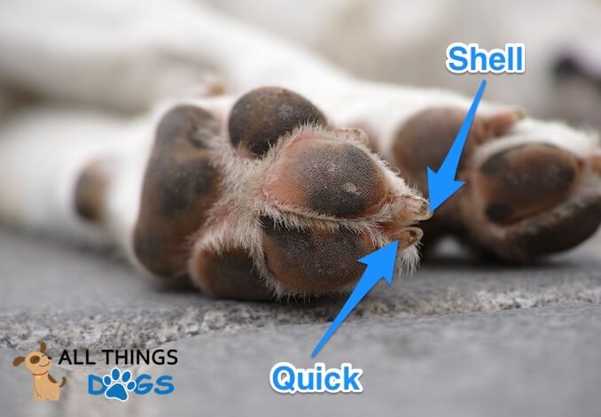
The nail consists of a harder outer shell which is usually either pink/white or black. Inside of this shell is a soft cuticle which is known as the quick; this has a blood supply and a nerve.
You will notice the quick more easily in light colored nails; it is the pinkish area.
When nails are trimmed, if the quick is caught; it will bleed and cause pain.
Most dogs if walked regularly will keep their nails down. If you are concerned about the length of your dog’s nails, speak with your veterinarian. They may even show you how to safely trim them.
Dog Skull Anatomy
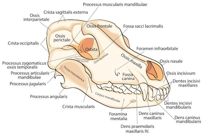
The main function of the skull in dogs and humans alike is to protect the brain. The interesting thing about dog’s skulls is that they can be different shapes and sizes.
Most breeds are categorized into the following dolichocephalic, mesocephalic and brachycephalic:
- Dolichocephalic – long headed such as the King Shepherd
- Mesocephalic – skull shape and size somewhere in the middle like the Golden Retriever
- Brachycephalic – broader, wide-skulled dogs like the Chihuahua
Interestingly, one studied demonstrated that head shape and size correlated with specific behaviors:
- Brachycephalic dogs tend to show more interest in humans and appear to be more defensive.
- Dolichocephalic breeds are less likely to engage in play but they are also less easily startled and appear to be more resilient in stressful situations.
Dog Teeth
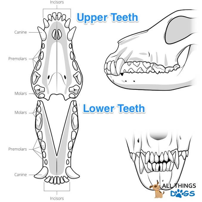
Also inside of their skull are teeth… a grand total of 42 teeth include 10 molars, 16 pre-molars, 4 canines and 12 incisors when fully grown.
Not surprisingly they have all evolved to serve a particular function:
- The canines are designed to grasp and tear.
- The incisors are used for cutting or shearing into food.
- The pre-molars have a flat biting surface – ideal for crushing.
- The molars are the largest teeth, having a flat surface which is ideal for grinding, crushing and chewing food.
But they don’t have 42 teeth to start off with. Puppies are born toothless; soon growing their 28 puppy teeth. Those razor sharp teeth dig into your fingers. You’ll need to train your puppy to not bite.
At around 3 months old, these puppy teeth fall out due to the adult teeth pushing through. You may find puppy teeth around your home, but many puppies swallow them.
The tooth consists of a crown, which is usually covered by enamel. Enamel is the hardest substance found in the body. The crown meets the root which is encapsulated by the alveolar bone; known as the tooth sockets found in the jaw bone.
Dog Muscle Anatomy
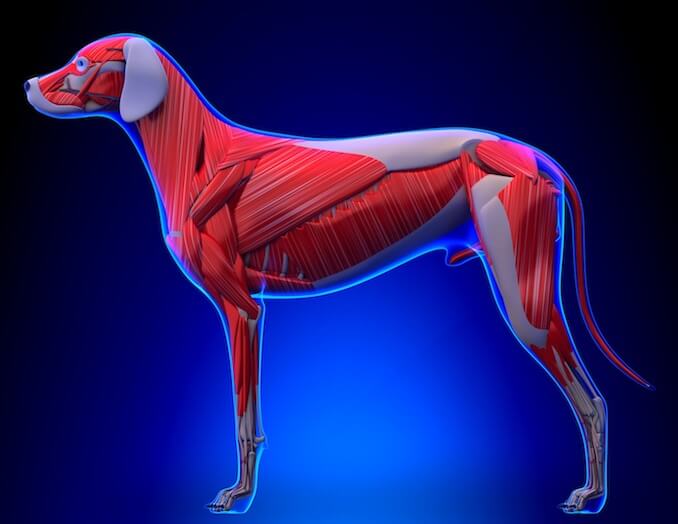
Muscles enable us to move. They stabilize our joints and maintain our posture; this is exactly the same for dogs.
Muscle fibers receive electrical impulses from the brain through the central nervous system which tells it whether to contract or elongate; therefore creating movement.
There are a number of different types of muscles.
Skeletal muscles are connected directly to bones by tendons (elastic type fibers).
Visceral muscles are found inside organs such as the stomach, intestines and blood vessels.
You may notice muscle atrophy (loss) in dogs who have an injury or developmental issue. Dogs with hip dysplasia or cruciate damage will often have muscle loss in their affected legs.
Injury or over-exertion can often cause muscle spasms, which appear as a localized twitch. This is caused by an interruption in the normal muscle contraction. They can be involuntary or sometimes caused by touch.
Dog Ear Anatomy
Another part of the dog’s head which can come in all shapes and sizes are their ears. Some are floppy, some are tall and pointy and some are just mud and water magnets… Spaniel owners will be nodding in agreement!
While a dog may totally ignore you in search of that squirrel, his hearing is actually pretty incredible. A human’s range of hearing is between 64Hz to 23kHz. A dog’s range is 67Hz to 45kHz; a much bigger range.
Their ears have a similar anatomy to that of a human. The outer ear (the bit you see covered in skin and fur) includes the pinna and also the canal (i.e. ear canal). The pinna gathers sound waves, sends them down the ear canal to the ear drum.
The ear drum makes up the middle ear along with those three little bones; the hammer, anvil and stirrup.
We then have the inner ear; this includes the cochlea and vestibular system (which supports balance).
Female vs Male Dog Anatomy
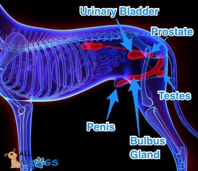
Not only are females generally smaller than males in the dog world; they have a few other differences too.
The male has what’s known as a bulbus glandis. This is what’s responsible for the tie that occurs during mating.
During copulation, the bulbus glandis engorges with blood which makes it swell.
This swelling locks it into the female vagina, demonstrating the typical “knot” or “tie”.
When you understand the biology of what happens you can see why trying to separate a male and female during the tie is inadvisable.
As in humans, their testes produce sperm and testosterone. Their prostate produces fluid that helps transport and support sperm. Some of its secretions also have antibacterial properties.
What About The Females?
The female reproductive tract includes the ovaries, uterus, vagina, vulva and mammary glands. Ovaries release eggs and produce reproductive hormones.
The uterus is Y shaped in our four legged friend. You may have heard it being called the uterine horn due to its shape.
When pregnant, the puppies arrange in both horns.
At the bottom of the uterus is the cervix, which leads to the vagina. The vulva protects the entrance to the vagina.
The mammary glands are where you will find puppies suckling milk from their Mom. A dog usually has 5 pairs of mammary glands.
Neutering in males removes both testicles so they are unable to produce sperm or testosterone. Spaying female dogs removes their ovaries and uterus, rendering her unable to become pregnant. Neutering is generally tolerated well with few long term side effects.
Summary
Despite the range of dog breeds, they really all have the same anatomy.
A skeletal system, powerful muscles for movement, ears for listening (or not as the case may be) and teeth for grinding and tearing.
Whilst males and females have their obvious reproductive differences, it is still incredible that within a breed they look identical.
What is interesting is how different shapes and sizes can be a factor in behavior, especially that brachy breeds tend to show more interest in humans and dolicocephalic breeds are less easily startled and also how different breeds have more fast twitch muscle fibers than others.
Having a basic knowledge of dog anatomy helps us understand our four legged friends and proves why despite their reputation as speed freaks; greyhounds are actually incredibly lazy.


My dog caught his shoulder while running up the steps. It was more like a trip. A few days later he started carrying his leg, nothing is broken we have had xray done. His leg muscle is swollen and he won’t use his leg, any advice?
Hi Bryan, you mention that you have had an x-ray done, what advice did your Veterinarian give? It’s important to follow the recommendation of your vet as they have seen your dog and understand the injury. If you are concerned your dog’s condition is worsening, pop to see your Vet for further investigation or advice. We hope your pooch recovers soon.
Can you tell me if dogs have some sort of glands on their backs that can secrete a little fluid from time to time? I have 3 different dogs that seem to have this happen at times. The liquid appeara to be clear but smells horribly fishy. Can you enlighten me on this matter?
Yes, you are correct. Dogs have anal glands. They are scent glands located between the muscles that make up the rectum. When working correctly, they are expressed when your dog poops. It’s a way to tell other dogs they’ve been there. Some dogs can suffer with impacted anal glands, meaning they no longer express. This can be for a range of reasons, often related to diet.
If your dog has a run of loose stools, there isn’t anything of substance to help express. Other reasons include allergies and infection. If a dog is having issues with their anal glands, they may scoot across the floor or excessively lick the area to try and express them themselves.
Groomers will sometimes offer a service to express them, but guidelines in many countries have advised against this as it can often cause future problems. The best port of call is your veterinarian.
My German Shepherd can’t sit properly. The part of her legs between the hock & feet are really tender. She’s a working dog so needs to be fit. Is there anything I can do for her or is it an immediate visit to the vets?
Hi Jeff – probably best to take her to the vets if it hasn’t gotten any better after 24 hours of rest.
My miniature poodle strained or sprained her left hind leg. She has been to the vet and x-rayed. At the same time, she had a bladder infection, and the x-ray showed crystals or stones in the bladder. She was on a special diet, antibiotics and anti-inflammatory meds. She is still on the anti-inflammatory and is slowly improving, but I am trying to understand what muscle or tendon was injured that would show up in the same x-ray as the bladder. Something in the hip, I assume. I have a follow-up appointment with a surgeon since my regular vet thought they would be able to interpret the x-ray better than she can. I have to way 2 more weeks for that appointment, and it is difficult to keep this dog at rest because she is very active by nature. Any added information you can provide would be appreciated.
hi there I’m doing a research project on dog sledding and was wondering if huskies and malamutes have a slightly different anatomical system???
E.G. stronger heart, stronger muscles, slightly different skeletal system?
would be great to get an answer because I can’t find anything anywhere!! 🙂